Featured
- Get link
- X
- Other Apps
Heart Ultrasound Test
This can be used to enhance an echo contrast echo but is not necessary for all echocardiograms. An echocardiogram echo is a test that uses high frequency sound waves ultrasound to make pictures of your heart.
An echo test is an ultrasound of the heart.

Heart ultrasound test. It can lead to a quick. The device gives off a silent sound wave that bounces off the heart creating images of the chambers and valves. Echocardiography is generally painless.
A sedative is not needed for an echo. The echocardiogram has been the most widely-used diagnostic test for heart disease for over 50 years. It uses high-frequency sound waves to produce images of the hearts valves and chambers so that doctors can see how your heart is functioning.
An echocardiogram echosound cardheart gramdrawing is an ultrasound test that can evaluate the structures of the heart as well as the direction of blood flow within it. An echocardiogram is a test that uses ultrasound to show how your heart muscle and valves are working. In a traditional echocardiogram the patients heart is ultrasounded to create a picture of the heart allowing medical professionals to assess the.
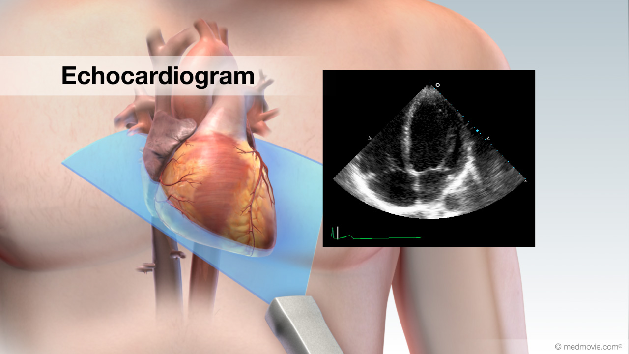
During an echocardiogram a doctor will generate a real-time image of the heart. The sound waves make moving pictures of your heart so your doctor can get a. The test monitors ultrasound high-frequency sound waves which are projected through the chest and bounce back to create an image of your heart.
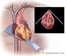
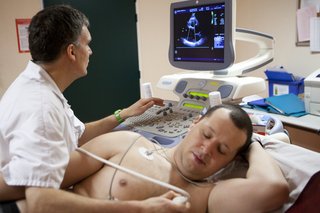
During an echo test ultrasound high-frequency sound waves from a hand-held wand placed on your chest provides pictures of the hearts valves and chambers and helps the sonographer evaluate the pumping action of the heart. A cardiac sonographer will move a hand-held device called a transducer over the chest area. Echo is often combined with Doppler ultrasound and color Doppler to evaluate blood flow across the hearts valves.
High-frequency sound waves are sent to the heart and transmitted back to the ultrasound machine as live moving images. An echocardiogram echo is an ultrasound test that images the moving heart. Ultrasound imaging procedure used to assess cardiac function.
The echocardiogram can show all four chambers of the heart the heart valves the blood vessels entering and leaving the heart and the sack around the heart. Echocardiography is an ultrasound test that uses sound waves to examine the hearts structure and motion. Echocardiography allows doctors to visualize the anatomy structure and function of the heart.
The test is useful for diagnosing and monitoring heart problems and creating treatment plans. The patient lies motionless while a technician moves a device over the chest. The test is also called echocardiography or diagnostic cardiac ultrasound.
Injection of a dye is not involved in a regular echo.
 Echocardiogram Vs Ekg Explained By A Cardiologist Myheart
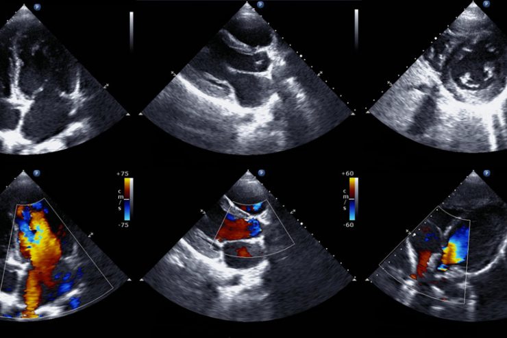 What To Expect During An Echocardiogram Upmc Healthbeat
 Why Would I Need A Heart Ultrasound Mayfair Diagnostics
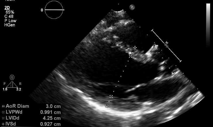 What Does An Echocardiogram Show Myheart
 Having An Echocardiogram What You Need To Know Upmc Healthbeat
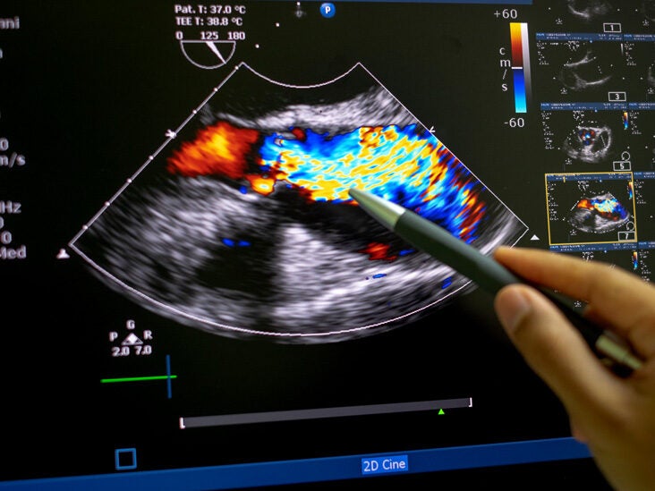 When Does Medicare Pay For Echocardiograms Cover Rules And Costs
 A Cardiologist Answers What Is An Echocardiogram And Why Do I Need One Health Essentials From Cleveland Clinic
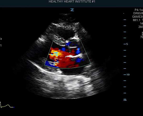 Echocardiogram Healthy Heart Institute
 Cardiac Ultrasound Apical View Sonosite Inc Youtube
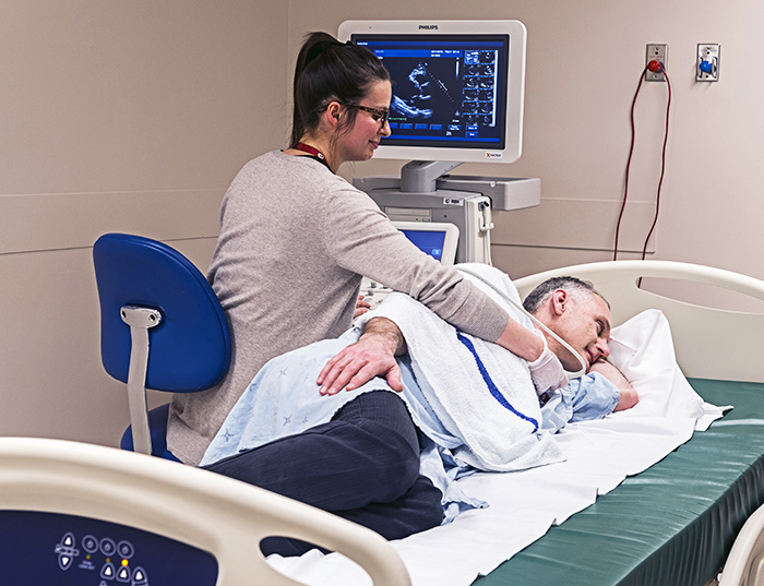 Echocardiogram Ottawa Heart Institute
 Echocardiography Senior Patient Undergoing A Heart Ultrasound Scan Stock Photo Alamy
/1745246_color1-5b9fd36cc9e77c002ce2dafa.png) Echocardiogram Uses Side Effects Procedure Results
Echocardiogram Uses Side Effects Procedure Results
Comments
Post a Comment